
- October 23, 2026
- DrnikhilK
- 9:07 am
What Is Meningioma?
What Are the Common Symptoms of Meningioma?
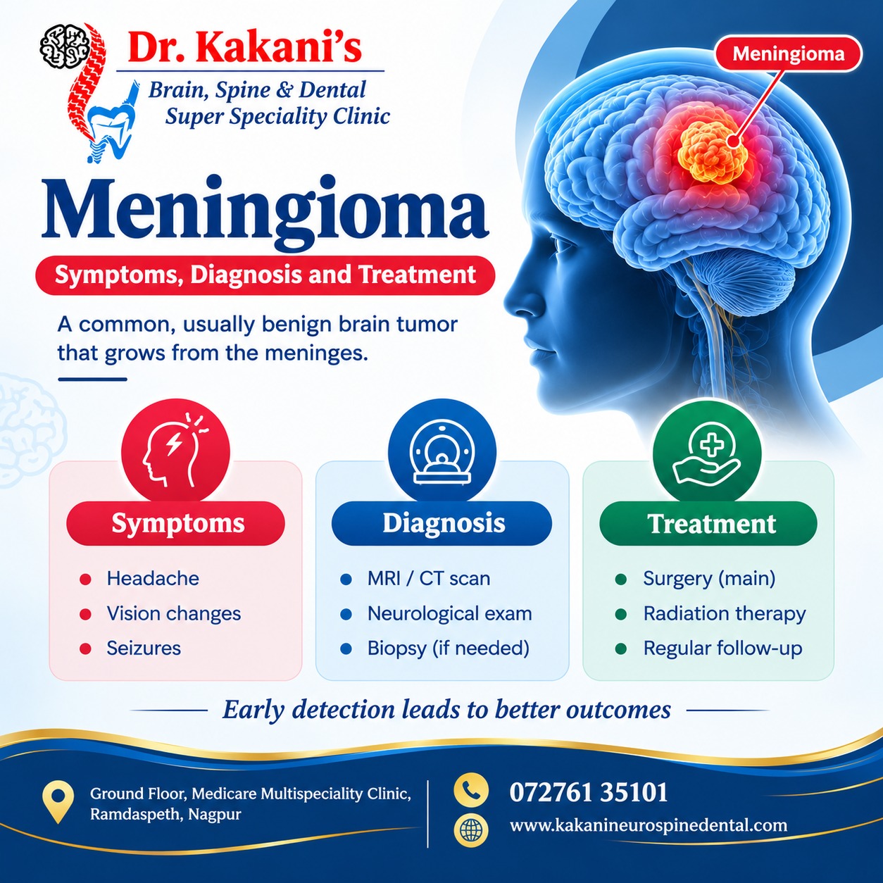
Meningioma symptoms vary depending on the tumour’s location, size, and pressure on nearby nerves or brain tissue. Some people may not experience any symptoms, particularly when the tumour is small.
Common meningioma symptoms include:
Persistent headaches: Headaches that are new, worsening, or different from usual.
Seizures: Episodes of abnormal electrical activity in the brain.
Vision problems: Blurred vision, double vision, or reduced visual function.
Hearing difficulties: Changes in hearing or ringing in the ears in some cases.
Weakness or numbness: Symptoms affecting the arms, legs, or one side of the body.
Memory and concentration problems: Difficulty focusing, remembering information, or thinking clearly.
Balance and coordination issues: Difficulty walking steadily or coordinating movements.
Personality or behavioural changes: Changes in mood, behaviour, or mental functioning.
Symptoms should be assessed by a qualified medical professional, especially if they are persistent or progressively worsening.
What Causes Meningioma?
The exact cause of meningioma is not always known. These tumours develop when cells in the meninges grow abnormally.
Factors associated with an increased risk of meningioma include:
Age: Meningioma is more common in adults, particularly older adults.
Radiation exposure: Previous radiation treatment involving the head may increase risk.
Genetic factors: Certain inherited conditions, such as neurofibromatosis type 2, are associated with meningioma.
Hormonal influences: Some meningiomas have hormone receptors, although the relationship between hormones and tumour growth is complex.
Having a risk factor does not necessarily mean a person will develop meningioma.
How Is Meningioma Diagnosed?
Accurate diagnosis is essential for selecting the right meningioma treatment. A specialist may recommend the following investigations based on symptoms and clinical findings.
1. Neurological Examination
A neurological examination helps assess muscle strength, reflexes, balance, coordination, vision, and other functions that may be affected by a tumour.
2. MRI Brain Scan
Magnetic Resonance Imaging (MRI) of the brain is commonly used to identify a meningioma, evaluate its size and location, and assess its relationship with surrounding structures.
3. CT Scan
A CT scan may help identify calcification, changes in nearby bones, or other features that support diagnosis and treatment planning.
4. Biopsy and Histopathological Examination
When surgery or tissue sampling is performed, laboratory examination of the tumour helps determine its type and grade. A biopsy is not required in every case.
5. Follow-Up Imaging
For small, asymptomatic meningiomas, periodic MRI scans may be recommended to monitor tumour growth and identify changes that require treatment.
What Are the Treatment Options for Meningioma?
Meningioma treatment is individualized according to the tumour’s characteristics, symptoms, and the patient’s overall health.
1. Active Surveillance
Small meningiomas that are not causing symptoms and do not show concerning growth may be monitored through regular clinical assessments and MRI scans.
2. Meningioma Surgery
Surgery may be recommended when the tumour causes symptoms, grows over time, produces pressure on important structures, or requires tissue diagnosis. The surgical approach depends on the tumour’s location and relationship with nearby nerves and blood vessels.
The main goals are to remove as much of the tumour as safely possible, relieve pressure, and preserve neurological function.
3. Radiation Therapy and Radiosurgery
Stereotactic radiosurgery or fractionated radiation therapy may be considered for selected meningiomas, including some small tumours in challenging locations, residual tumours after surgery, or tumours that recur. The most suitable approach depends on tumour size, location, grade, and previous treatment.
4. Follow-Up Care
Regular follow-up is important after treatment because some meningiomas may recur or continue to grow. Follow-up schedules depend on tumour grade, treatment results, and individual risk factors.
Recovery After Meningioma Treatment
Recovery varies depending on the tumour’s location, size, treatment method, and the person’s general health.
After meningioma surgery, the care plan may include:
Monitoring neurological function and general recovery.
Follow-up appointments and MRI scans.
Medication when prescribed for specific symptoms or complications.
Physiotherapy or rehabilitation if movement, balance, or strength is affected.
Gradual return to daily activities according to medical advice.
Patients should follow their treating specialist’s instructions and attend all scheduled follow-up appointments.
When Should You Consult a Specialist?
Consult a medical professional if you experience persistent or worsening headaches, unexplained seizures, vision changes, weakness, balance difficulties, or other new neurological symptoms.
Seek emergency medical care for a first seizure, sudden weakness, sudden vision loss, severe sudden headache, or new confusion.
Early assessment can help identify the cause of symptoms and determine whether further investigations or treatment are necessary.
Meningioma Evaluation and Treatment with Dr. Nikhil Kakani
Consult Dr. Kakani's Clinic
Dr. Kakani’s Brain, Spine & Dental Super Speciality Clinic
Appointment: 07261 35101
Website: www.kakineurospinedental.com
Address: Ground Floor, Medicine Multispeciality Clinic, Ramdaspeth, Nagpur.