- November 2, 2026
- DrnikhilK
- 12:37 pm
What Is a CT Scan for Brain Tumor Diagnosis?
A Computed Tomography (CT) scan uses X-rays and computer technology to create detailed cross-sectional images of the brain.
During the scan, the machine captures multiple images from different angles. The computer then combines these images to create a detailed view of the brain and surrounding structures.
Doctors may recommend a brain CT scan to:
Detect abnormal growths or suspected brain tumors.
Identify bleeding inside the brain.
Examine swelling and changes in brain structures.
Evaluate skull abnormalities and bone involvement.
Assess emergency neurological conditions.
Moreover, a CT scan takes only a few minutes, making it useful when doctors need quick results.
What Is an MRI Scan for Brain Tumor Diagnosis?
Magnetic Resonance Imaging (MRI) uses strong magnetic fields and radio waves to produce detailed images of the brain.
Unlike CT scans, MRI does not use ionising radiation. It provides excellent soft-tissue detail, helping doctors examine abnormal brain tissue and understand how a tumor affects nearby structures.
Doctors commonly recommend an MRI brain scan to:
Detect small or difficult-to-identify brain tumors.
Determine the exact location and size of a tumor.
Examine surrounding brain tissue and swelling.
Identify tumors that have spread from other parts of the body.
Plan brain tumor surgery and further treatment.
Additionally, doctors may recommend a contrast-enhanced MRI. This scan uses a gadolinium-based contrast agent to make certain abnormal areas more visible.
CT Scan vs MRI for Brain Tumor Diagnosis: Key Differences
Although both imaging tests help identify brain abnormalities, they differ in technology, image quality and clinical applications.
| Feature | CT Scan | MRI Scan |
|---|---|---|
| Technology | X-rays | Magnetic fields and radio waves |
| Scan duration | Usually a few minutes | Usually 30–60 minutes |
| Soft tissue detail | Less detailed | More detailed |
| Radiation | Uses ionising radiation | No ionising radiation |
| Emergency diagnosis | Very useful | Less commonly used as the first emergency scan |
| Tumor evaluation | Helps detect abnormalities | Provides detailed tumor assessment |
| Bone examination | Excellent | Less detailed than CT |
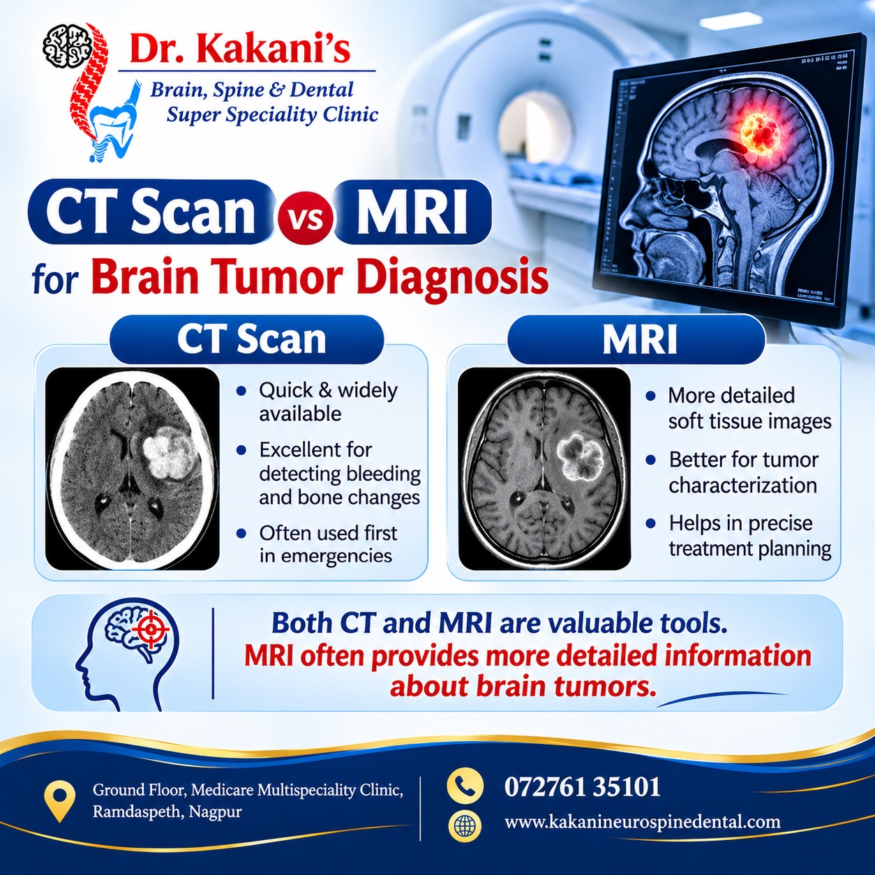
Therefore, MRI generally provides more detailed information about brain tumors, while CT remains valuable in emergency situations and for examining bones and acute bleeding.
Which Is Better for Detecting Brain Tumors: CT Scan or MRI?
In most cases, MRI is the preferred imaging technique for evaluating suspected brain tumors.
MRI provides clearer images of brain tissue and helps doctors identify the tumor’s location, size and relationship with important surrounding structures.
However, CT scans also have an important role in brain tumor diagnosis. Doctors may recommend a CT scan when:
A patient requires immediate emergency evaluation.
There is a suspicion of bleeding inside the brain.
An MRI is not suitable because of certain medical implants or other safety concerns.
Doctors need to examine nearby skull bones.
In some cases, doctors may recommend both CT and MRI scans to obtain complementary diagnostic information.
Why Is Contrast-Enhanced MRI Important?
Contrast-enhanced MRI helps doctors identify certain abnormalities more clearly.
During this procedure, a contrast agent is injected into a vein before or during the scan. It helps highlight areas where the blood-brain barrier is disrupted or abnormal blood vessels are present.
Furthermore, contrast-enhanced MRI can help doctors:
Identify tumor boundaries more clearly.
Assess tumor enhancement patterns.
Examine surrounding swelling.
Evaluate possible brain metastases.
Plan further investigations and treatment.
The doctor decides whether contrast is necessary based on the patient’s medical history, kidney function and diagnostic requirements.
Can a CT Scan or MRI Confirm the Type of Brain Tumor?
Imaging tests provide important information about a suspected brain tumor. However, a scan alone may not always confirm its exact type or grade.
Doctors evaluate the imaging findings along with symptoms, neurological examination and medical history.
In many cases, a biopsy or examination of surgically removed tumor tissue helps establish the definitive diagnosis. Laboratory analysis also helps identify the tumor’s specific characteristics and guide treatment decisions.
Therefore, accurate diagnosis often involves a combination of imaging and other medical investigations.
What Happens After a Brain Tumor Is Detected?
Once imaging tests identify a suspected brain tumor, the next step depends on its location, size, appearance and the patient’s overall health.
A neurosurgeon may recommend:
Further Imaging: Additional MRI sequences or specialised imaging may provide more information.
Neurological Evaluation: Doctors assess vision, speech, memory, coordination and muscle strength.
Biopsy: A tissue sample may be required to identify the tumor type.
Treatment Planning: Doctors evaluate suitable options, including surgery, radiation therapy or other treatments.
Follow-Up: Regular imaging helps monitor the tumor and assess treatment response.
Moreover, an individualised treatment plan helps doctors address the specific needs of each patient.
When Should You Consult a Neurosurgeon?
Certain neurological symptoms require medical evaluation, especially when they persist, worsen or appear unexpectedly.
Watch for symptoms such as:
Persistent or worsening headaches.
Repeated episodes of vomiting.
Seizures or unexplained fits.
Blurred or double vision.
Difficulty speaking or understanding language.
Weakness or numbness in the arms or legs.
Changes in memory, behaviour or balance.
However, these symptoms do not always indicate a brain tumor. They can occur due to several neurological conditions. Seek immediate medical attention for a first-time seizure, sudden weakness, sudden speech or vision changes, or a sudden severe headache.
Consult Dr. Kakani's Clinic
Dr. Kakani’s Brain, Spine & Dental Super Speciality Clinic
Appointment: 07261 35101
Website: www.kakineurospinedental.com
Address: Ground Floor, Medicine Multispeciality Clinic, Ramdaspeth, Nagpur.